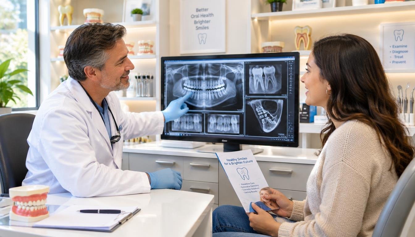
A routine dental exam can miss changes between teeth or beneath the gumline, which is why a dentist may also recommend dental X-rays. The right image depends on your needs, not simply on the fact that you are attending a checkup.
That can matter during routine dental visits, especially when hidden oral health concerns are hard to spot by sight alone. New dental patients may also need to share past images or ask why an exam is part of their care.
This guide explains what different exams can show, why a dentist may recommend them, and what questions you can ask. Ask what the image is intended to check if you are unsure why it is part of your visit.
Key Takeaways
- Dental images help assess areas that a visual exam cannot fully show.
- The image you need depends on your symptoms, history, and dental exam.
- An image alone cannot confirm every tooth problem or set a treatment plan.
- Ask whether a recent image can answer the same clinical question.
- Tell your dentist about symptoms, pregnancy, or concerns about radiation.
What Can Dental X-Rays Reveal Beyond Cavities?
A dental X-ray can reveal decay between teeth and changes around a root that a visual exam may not show. These hidden findings can help your dentist check cavities, gum disease, and other concerns.
Radiographs cannot guide diagnosis or treatment planning on their own. Your dentist also considers symptoms, an exam, and tests of the tooth’s pulp.
A dental X-ray can show signs around a tooth, but it cannot decide by itself whether you need a root canal.
Hidden changes in teeth, roots, and jawbone
Images can reveal decay between teeth or beneath fillings and other restorations. They can also show bone loss linked to gum disease, impacted teeth, and changes near a root that may suggest an abscess.
Dentists may find cysts, some tumors, or other changes in the jaw. Images can also help plan treatment and check healing after care, such as an extraction or other procedure.
What images can and cannot show about pulp problems
Pulpitis means inflammation of the pulp, the inner tissue containing nerves, blood vessels, and connective tissue. Brief sensitivity to cold or sweets can occur with reversible pulpitis.
Sensitivity to heat, cold, or sweets lasting more than a few seconds can occur with irreversible pulpitis. Electric pulp testing checks whether pulp tissue is alive; no response may point to pulp necrosis.
Heat or cold tests and tapping the tooth can help assess pulpitis. Pain when the dentist taps a tooth can be a sign of irreversible pulpitis.
An image may show tooth defects or infection signs, but it does not directly prove nerve damage. Your dentist combines symptoms, examination, imaging, and pulp tests before deciding what care may help.
Tell the dentist about lingering temperature sensitivity or pain on tapping, since those symptoms help guide the examination.
Which Type of Dental X-Ray Shows Each Area?
A bitewing is suited to checking between back teeth, while a periapical image follows one tooth from crown to root. Each type gives your dentist a different view, from a few teeth to the jaws.
The image choice depends on the area and question being examined. The table compares five types dental X-rays and their common uses.
Bitewing and periapical images inside the mouth
Bitewings show the upper and lower tooth crowns in one section of the mouth. Dentists often use them to check for decay between teeth and changes in bone near the teeth.
Periapical X-rays show a whole tooth from crown to root tip. They can help assess a tooth with pain or a concern near its root.
| Type | Location and view | Image format | Common use |
|---|---|---|---|
| Bitewing | Upper and lower crowns in one section | 2D, inside mouth | Decay between teeth |
| Periapical | One tooth, crown to root tip | 2D, inside mouth | Root and tooth concerns |
| Full-mouth series | Teeth throughout the mouth | Set of 14-21 images | Broad review of teeth and roots |
| Panoramic | Teeth, upper and lower jaws, and nearby structures | Broad 2D view | Jaw and treatment planning |
| CBCT | Selected area of teeth and jaw | 3D view | Complex treatment planning |
Full-mouth series and occlusal images
A full-mouth series combines images of the teeth and roots. It usually contains 14-21 images, rather than one wide view of the whole mouth.
Occlusal images show a broad section or arch. A dentist may use one to review a larger area of the mouth in a single image.
Panoramic, cephalometric, and cone beam images
A panoramic X-ray captures the teeth, upper and lower jaws, and nearby structures in one broad 2D image. Its rotating arm typically moves around your head for 12-20 seconds while it captures the image.
Panoramic images can help plan dentures, braces, extractions, and implants. They may show impacted teeth, jaw cysts, jaw tumors, and some jaw disorders, but they give less detail about individual teeth and soft tissues than images taken inside the mouth.
A cephalometric image gives a side view for orthodontic planning. Cone beam CT (CBCT) creates 3D views for complex planning, such as assessing a selected area before treatment.
For a child aged 6-12, a panoramic image may be useful when the dentist needs to review permanent-tooth eruption or orthodontic concerns.
Dental X-Rays Explained: Types, Benefits, Safety, and Uses: How Often Are They Needed?
Higher-risk patients may need bitewing images every 6 months, while lower-risk adults may need them every 18-24 months. Those intervals are examples, not a schedule for every patient.
The American Dental Association recommends choosing images based on each patient’s needs, not a fixed schedule. Age, symptoms, oral-health history, and disease risk all help guide the timing.
Children may need imaging to assess how their teeth and jaws are developing. Your dentist may also consider changes in symptoms or oral health since your last exam.
Risk factors that may lead to more frequent bitewings include:
- Frequent cavities
- Active gum disease
- Dry mouth
- Diabetes
- Orthodontic treatment
So, x-rays every 6 months may fit some higher-risk patients, but not every image or person. A lower-risk adult may need bitewings less often, while a new symptom can call for an image sooner.
Ask your dentist which personal risk factors are changing your recommended interval.
How Safe Are Dental X-Rays?
Dental X-rays use a low radiation dose, and digital systems typically reduce exposure by 70-80% compared with traditional film. Digital imaging can lower exposure while still giving the dental team useful images.
Effective dose is a calculated estimate of the potential biological effect of radiation exposure, expressed in sieverts. It is not a direct measurement of radiation in your body.
Understanding dose and digital imaging
A full set of dental X-rays has an approximate effective dose of 0.005 millisieverts. The FDA lists an average adult effective-dose range of 0.005-0.016 mSv for a dental X-ray.
| Measure | Amount | What it describes |
|---|---|---|
| Approximate effective dose for a full set | 0.005 mSv | Estimated radiation risk |
| FDA average adult dental X-ray effective-dose range | 0.005-0.016 mSv | Estimated dose range |
| Digital-system exposure reduction compared with film | 70-80% | Typical exposure reduction |
Effective dose helps compare the potential effects of different exposures. It does not predict an individual patient’s health outcome.
How dental teams limit exposure
Dental teams follow ALARA, which means “As Low As Reasonably Achievable.” Imaging should have a clear clinical reason and use suitable exposure settings.
Teams can limit exposure by using a fast image receptor, aiming the beam at the image receptor where possible, and choosing proper settings. These steps help keep exposure as low as reasonably achievable.
Pregnancy or a concern about radiation is a reason to discuss the specific need with your dentist, rather than skip necessary care. Your dentist can explain how the proposed image may guide treatment.
If you are concerned about exposure, ask what the image will help diagnose and whether an existing image can answer the same question.
What Happens During a Dental X-Ray Appointment?
Most dental X-ray appointments involve positioning a sensor or machine, holding still briefly, and repeating the process only if additional views are needed. Good positioning helps the team capture a clear image.
Stay as still as you can while the image is taken. The dental team will place the sensor or guide you to the right position for the selected view.
Positioning and taking the image
For images taken inside the mouth, a team member places a small sensor beside or between your teeth. You may need to close gently on a holder while the image is captured.
For extraoral images, you stand or sit at a machine. The team positions your head and asks you to stay still as the machine captures the view.
You can ask these questions during the visit:
- Which area will this image show?
- Can an existing image be used?
- How should I hold still or position my mouth?
- How can I get a copy of the image?
Requesting and transferring existing images
When changing offices, ask your previous dentist to send your images directly to the new office. You can also request copies for your own records.
Images from the past 12-24 months may transfer to a new office, depending on clinical need and image quality. A newer image may not answer the current question if your symptoms or treatment needs have changed.
HIPAA generally protects your right to access dental records, including images. Office procedures apply to requests, so ask how to submit one and where the office can send the files.
Tell the dental team if sensor placement causes discomfort so they can help adjust it.
What Should You Check About Coverage and Existing Images?
Dental plans may cover bitewings annually or every 12-18 months, while panoramic images may be covered once every 3-5 years. Plan limits can affect payment, but they do not decide what care you need.
Coverage depends on your plan, the reason for imaging, and its frequency rules. Advanced imaging such as CBCT may face different review rules and may fall under dental or medical insurance, depending on the service.
A plan’s coverage limit is not a clinical schedule. Your dentist bases an imaging recommendation on your care needs.
Read your benefit documents for covered services, limits, and any preauthorization rules. Ask the office for a pre-treatment estimate when appropriate, but remember that an estimate does not guarantee payment or benefits.
Confirm the exact benefit and any preauthorization requirement with your plan before relying on an estimated payment.
Conclusion
The most useful dental X-ray is the one that answers a specific clinical question. A clear reason for imaging helps you understand how it supports your oral health and care.
At your next visit, ask what the image will help the dentist evaluate and whether an existing image can serve that purpose.